


A venogram is an imaging test that shows the inside of your veins. Often, dye (contrast) is put into a vein, and pictures are taken to see whether a vein is narrowed, blocked, or leaking backward. It is the most accurate way to check how a vein is actually working.
There is more than one kind of venogram, and knowing which one you are booked for changes what to expect. This guide covers the types, why your doctor might order one, and what the day itself is like.
I am an interventional radiologist in Toronto and perform these procedures regularly, so this is written the way I explain it to my own patients.
Types of venogram
People often arrive unsure which test they are having. There are four main kinds, from the simplest to the most detailed.
CT venogram (CT venography). Dye goes in through a regular IV in your arm, and a CT scanner takes the pictures. It takes minutes, needs no sedation, and you go home right away.
MR venogram (MR venography). Similar idea, but using an MRI scanner instead of X-rays, so there is no radiation. It is particularly good at showing the pelvic and ovarian veins. It takes longer than CT and requires lying still inside the scanner. At our hospital we often use non-contrast MR venography, which means no IV and no dye at all.
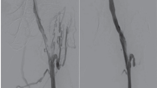
Central venogram (through an IV in your arm). This one sits between a scan and a full catheter procedure. Dye is injected through an ordinary IV in the crook of your elbow (the same kind used for bloodwork) while a live X-ray camera watches it travel up your arm, past your shoulder, and into the large veins in your chest. Nothing is threaded into the vein. It is quick, needs no sedation, and shows whether the big central veins draining your arm are open, narrowed, or blocked.
Catheter venogram. This is the detailed one, and what most people mean by “venogram.” A thin tube called a catheter is placed directly into a vein through a small needle poke. Dye is injected right where it matters, and live X-ray pictures show blood moving in real time. Pressure inside the vein can also be measured.
These tests are complementary rather than competing. A CT or MR venogram is often the first look, mapping the veins from the outside. A catheter venogram is the next step when that picture is unclear, because it shows blood actually moving and can measure pressures, things a scan cannot do.
Why your doctor might order a venogram
The common reasons fall into a few groups.
To find or assess a blood clot. When a deep vein thrombosis (DVT) is suspected, ultrasound is almost always the first test. It’s quick, safe, no dye. A venogram is not the usual way to diagnose a clot. It becomes useful in the more complicated situations: when a clot sits high in the pelvis where ultrasound sees poorly, when symptoms persist despite treatment, or when an older clot has left the vein narrowed and scarred. A venogram may also reveal why a clot formed. For example, a vein squeezed by an overlying artery, which is a treatable cause of recurrent left-leg clots.
To check the pelvic and leg veins. Leg swelling, heaviness, or pelvic pain can come from a vein that is compressed or leaking backward. A venogram shows this directly. If this is your situation, I have written a separate guide to what a pelvic or iliac venogram involves.
To check the veins of the arm, shoulder, and chest. An upper-extremity or central venogram looks at the veins draining your arm. Common reasons include unexplained arm swelling, planning or troubleshooting dialysis access, problems with a central line or port, checking that a vein is open before a pacemaker or defibrillator lead is placed, or suspected narrowing where a vein passes between the collarbone and first rib.
To plan treatment. When a narrowing or blockage has already been found, a venogram confirms it precisely and shows exactly where it is, so that treatment can be planned properly.
How Do I Prepare for a Venogram Procedure?
Before your test:
- Your doctor may ask you not to eat or drink for a few hours.
- Tell your care team if you’re on any medications.
- Tell them about any allergies, especially to contrast dye or iodine.
- If you have kidney issues, let them know. Some dyes can affect kidney function.
- If you might be pregnant, be sure to tell your doctor.
Wear loose, comfortable clothing and leave jewelry at home (metal can interfere with the X-rays).
What Happens During the Venogram Procedure?
Most often, I perform venograms for iliac vein compression, ovarian vein embolization and/or deep vein thrombosis. For these problems, I usually access the veins in the right upper arm (basilic vein) or the left groin (left common femoral vein/greater saphenous vein). “Access” is the process of creating a direct, reliable pathway into the vein.
Here’s a step-by-step of what you’ll likely experience:
- You’ll lie down on an X-ray table.
- We clean the skin around the vein we will access.
- A numbing medicine is given where we obtain access. You might feel a quick pinch and a burning sensation. A small tube is then placed in the vein.
- The contrast dye is slowly injected. You may feel warm or flushed, but it shouldn’t hurt.
- As the dye moves through your veins, an X-ray machine takes moving pictures (like a video).
- You might be asked to hold your breath or stay very still for a moment to get clear images.
- When the pictures are finished, the dye is flushed out, and we remove the small tube and press on the spot to close the small hole vein.
How long does a venogram take?
The catheter venogram itself usually takes 30 to 60 minutes on the table. If a narrowed vein is treated during the same visit, with a balloon and often a stent, add roughly 30 to 45 minutes.
The full hospital visit is longer than the procedure. Allow a few hours in total: check-in and preparation beforehand, then one to two hours of rest afterward while the puncture site seals. Almost everyone goes home the same day.
A CT venogram is much quicker. The scan itself takes only a few minutes, and there is no recovery period.
Venogram aftercare: what to do afterward
After a catheter venogram you rest for one to two hours while the small opening in the vein seals itself. A nurse checks the site and your vital signs. Most people go home the same day.
For the rest of that day:
- No driving (if you had sedation, this applies for the full day)
- Take it easy. No heavy lifting or hard exercise
- Drink extra fluids to help clear the dye from your system
- Keep the puncture site clean and dry
Over the next few days, a bruise at the needle site is normal and often looks worse before it fades. Most people are back to normal activity within about 24 hours.
Call the clinic or seek care if you notice: bleeding or a rapidly growing lump at the puncture site, a leg that becomes much more swollen or painful, or fever, chest pain, or trouble breathing.
Is a venogram dangerous? Risks and safety
A venogram is a low-risk procedure. The most common issue is minor bruising or tenderness at the puncture site, which settles within a few days. Serious complications, significant bleeding, injury to the vein, blot clots, or an allergic reaction to the contrast dye, are rare. The radiation dose is low and focused.
Two situations deserve a conversation beforehand: reduced kidney function, because the dye is cleared by the kidneys, and a previous reaction to contrast, which can usually be managed with pre-medication. Your doctor will talk through the risks with you and make sure the benefits outweigh them.
If a problem is found
Because the catheter is already in the vein, a narrowing or blockage found during a catheter venogram can sometimes be treated in the same visit rather than at a second appointment. Whether that is likely in your case is discussed with you beforehand, based on your symptoms and previous imaging. Nothing that happens should be a surprise.
Treatment is its own topic, and I have covered it separately: see iliac vein stent recovery, week by week if a stent is part of your plan.
What the results will tell you
The results help answer questions like: are there any blood clots? Is blood flowing normally through your veins? Is there a blockage or narrowing that needs treatment? Your doctor will walk through the findings with you and explain the next steps.
Venogram: Frequently Asked Questions
No, a venogram is not considered surgery. It is a minimally invasive, image-guided procedure performed through a needle puncture in a vein. There is no incision, no stitches, and in most cases you go home the same day.
The distinction matters for practical reasons: because it is not surgery, a venogram does not usually require general anesthesia, an operating room, or an overnight stay. It is performed by an interventional radiologist in a procedure suite using X-ray guidance, typically with local freezing at the puncture site and, if needed, light sedation.
A venogram involves only mild, brief discomfort for most people. The most noticeable part is the sting of the local anesthetic at the puncture site, similar to having blood drawn. After that, you may feel pressure as the catheter moves and a warm, flushing sensation when the contrast dye is injected. The sensation is odd, but not painful.
If a blockage is treated during the same procedure (for example, if a narrowed vein is stretched with a balloon) that part might cause a deep, cramping ache lasting seconds to a minute. Your team will warn you before it happens, and sedation can be adjusted. Most patients rate the overall experience as far easier than they expected.
Recovery from a venogram is quick: most people rest for one to two hours afterward while the puncture site seals, go home the same day, and return to normal activities within 24 hours. A small bruise at the access site is common and fades within a week.
For the first day or two, avoid heavy lifting and strenuous exercise to protect the puncture site, and drink extra fluids to help clear the contrast dye. If a stent was placed during the procedure, the recovery is somewhat different. See my post on iliac vein stenting recovery.
A CT venogram is a noninvasive scan: contrast dye is injected through an ordinary IV in the arm and a CT scanner takes the pictures. A catheter venogram, the procedure described on this page, places a thin tube directly into the vein of interest, giving real-time images, pressure measurements, and the option to treat a problem immediately.
The two are complementary rather than competing. A CT venogram is often the first test to map the veins and look for compression or clot. A catheter venogram is the next step when the diagnosis needs confirming, often with intravascular ultrasound (IVUS), which images the vein from the inside, or when treatment such as angioplasty or stenting is planned in the same session.
Booked for a pelvic or iliac venogram specifically? See what a pelvic or iliac venogram involves, step by step. Physicians can find referral information for Ontario physicians here.


My daughter is 23, with probable NCS, and nMals for 4 years. NCS shows on her scans, CT, and CTA, a venogram laying showed nothing, but shows dramatically worse standing on ultrasound. Told we need an IVUS by last VS and we have to go out of country, how can we access in GTA. VS has dismissed us until IVUS done, so we are now stuck. We’ve heard you have lots of vascular compression experience.
Hi Sandra. Have you doctor send us a referral and we can review your file. We have access to IVUS at our center and can help sort out the next steps.